
What Is a Fundus Examination and Why Is It Done?
A fundus (dilated eye) examination magnifies the retina, optic nerve and vessels; a priority with diabetes, flashes or floaters. Message our Istanbul clinic on WhatsApp.
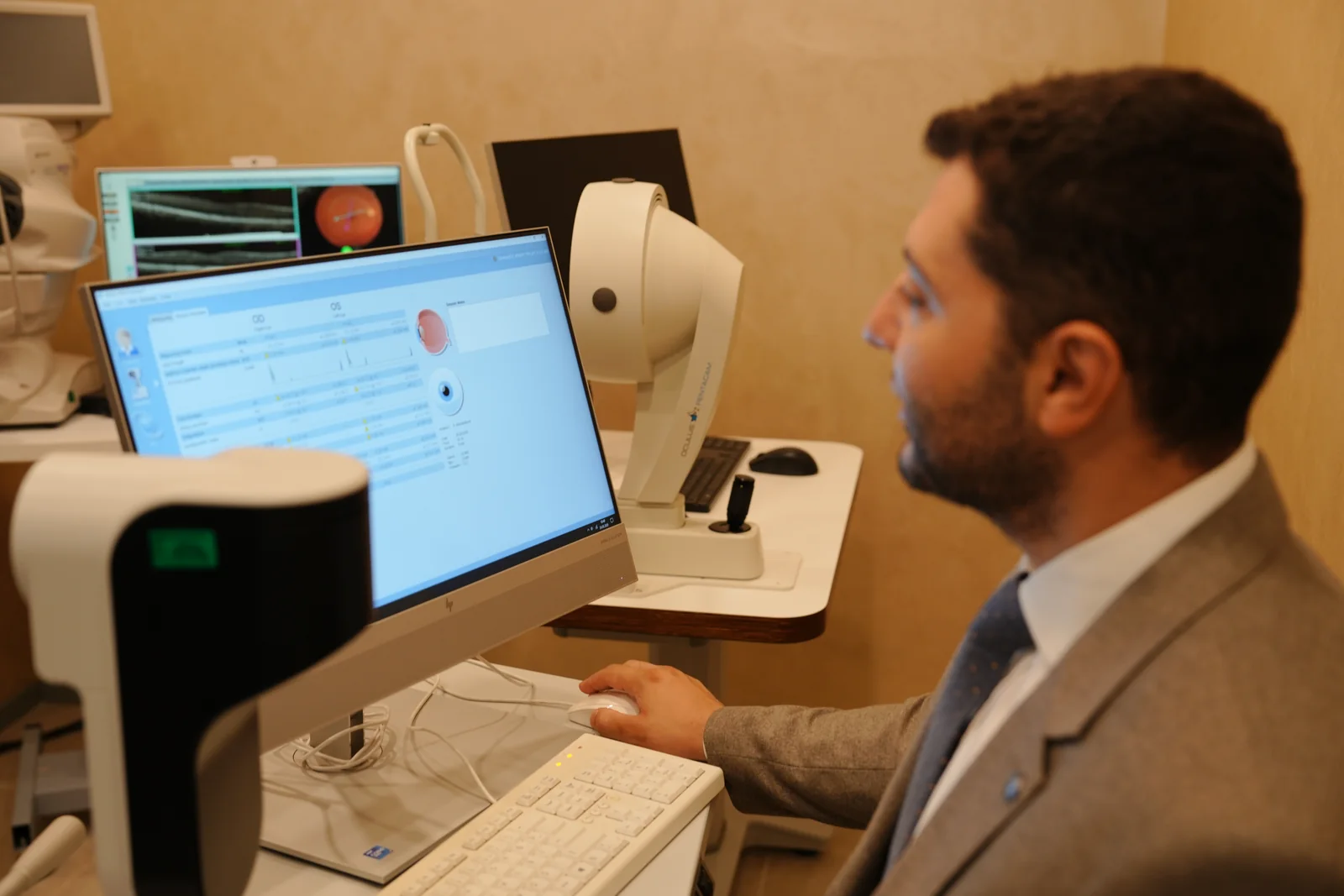

A fundus examination magnifies the retina, optic nerve and blood vessels at the back of the eye, usually after the pupils are dilated with drops. It is a priority for adults over 40, people with diabetes, those with a family history of glaucoma or retinal disease, and anyone noticing sudden flashes or floaters.
Written by Op. Dr. Anıl Kaya
What Is a Fundus Examination?
A fundus examination assesses the back of the eye — the retina, optic nerve and blood vessels crossing it — through a magnifying instrument. This area is not visible from the front of the eye; it becomes visible once the pupil is dilated, or with specialist lenses. It may extend a routine eye examination, or be requested on its own. Clinically called “fundoscopy”, it may involve a slit-lamp biomicroscope with a special lens, or a direct or indirect ophthalmoscope. Because it reveals structural eye health rather than visual acuity alone, it helps catch many eye conditions early. Even people with no complaints may be offered it during a routine eye examination, since eye health can change independently of visual acuity.
Quick Facts
A fundus examination looks at the retina, optic nerve and blood vessels at the back of the eye under magnification, usually after the pupil is widened with drops; significant discomfort is not expected. It is a priority for adults over 40, people with diabetes, those with high myopia, and anyone with a family history of glaucoma or retinal disease.
What Does a Fundus Examination Assess?
This examination looks at the retinal tissue, the macula, the optic nerve head, and the retinal blood vessels. Thinning, tears, bleeding or fluid build-up on the retina can be picked up here. Diabetes and high blood pressure can also leave their mark, as narrowing, bleeding or leakage in the retinal vessels. The colour and shape of the optic nerve head serve as a reference point for tracking glaucoma-related change. The peripheral retina is checked alongside the centre where possible, so findings not yet affecting central vision can still be caught; these may be followed up with imaging such as OCT.
Who Should Have a Fundus Examination?
A fundus examination is a priority for everyone over 40, anyone with diabetes, those with a family history of glaucoma or retinal disease, people with high myopia, and anyone noticing sudden flashes, floaters or a shadow across their vision. An ophthalmologist may also add it during a routine eye examination outside these groups. The recommended interval varies and tends to shorten as risk increases. Even adults with no major risk factors may be advised to repeat it occasionally, since changes at the back of the eye can progress without symptoms. The chart below outlines roughly how often checks are commonly recommended by risk group; the exact interval is set by the ophthalmologist.
Approximate recommended check frequency by risk group
Which Symptoms Call for a Fundus Examination?
Sudden loss of vision, floaters, flashes of light, or a fixed shadow in the visual field are situations where a fundus examination should be arranged as a priority. These can signal a problem at the back of the eye needing urgent assessment and should not be put off. Wavy or distorted vision, straight lines appearing bent, or sudden blurring in one eye call for similar attention. After an eye injury or eye surgery, or with a very high glasses prescription, an ophthalmologist may recommend a fundus examination even without symptoms. If any of these signs appear, an eye doctor should be seen promptly rather than waiting for a routine appointment.
How Is a Fundus Examination Performed?
Eye drops that widen the pupil are usually needed for a clear view of the back of the eye; these take around 20–30 minutes to take effect. The ophthalmologist then examines the retina and optic nerve using a special lens and a light source. Aside from a brief sensation of glare, the examination is not painful. While the drops are working, close-up tasks such as reading may feel harder and the eyes can become light-sensitive, so sunglasses and avoiding driving that day are advised. Including the wait, the whole visit rarely takes more than an hour; the examination itself is over within minutes.
- 1Pupil dilation. The ophthalmologist puts pupil-dilating drops into both eyes.
- 2Waiting period. A wait of around 20–30 minutes allows the drops to take effect.
- 3Viewing the back of the eye. The retina, macula and optic nerve are examined using a special lens and light source.
- 4Further tests if needed. If a finding needs a closer look, further imaging such as OCT may be arranged.
- 5Discussing the findings. At the end of the examination, the ophthalmologist discusses the findings with the patient and sets the recommended check interval.
What Do the Results of a Fundus Examination Mean?
If the findings are within normal limits, the next check is simply scheduled for later. When a change is found in the retina or optic nerve, the ophthalmologist assesses which condition it may be linked to — glaucoma, diabetic retinopathy, macular disease or a retinal tear — and arranges further tests if needed. Some findings only call for monitoring, while others need treatment sooner, depending on the type and extent of the finding. Comparing new findings with earlier records, where available, can help catch some slow-moving conditions early. The ophthalmologist explains what each finding means and the next step; further imaging may be requested if anything remains unclear.
Which Conditions Can a Fundus Examination Detect?
The findings most often picked up during a fundus examination are optic nerve changes linked to glaucoma, along with retinal conditions such as diabetic retinopathy, macular disease and retinal tears or detachment. Many of these can progress for some time without a symptom the patient would notice, giving a fundus examination real diagnostic value. Some findings are more common in older patients or those with long-standing diabetes, while a retinal tear is seen less often but needs urgent assessment. Some can only be picked up through a fundus examination; others need further imaging to confirm the diagnosis. The chart below groups possible findings by relative frequency, reflecting a general pattern rather than a fixed outcome for every patient.
Findings that can be detected during a fundus examination
If a Condition Is Found, What Are the Treatment Options?
When a finding turns up at the back of the eye, what happens next depends on the condition: glaucoma may call for medication, laser treatment or surgery, while retinal conditions may need targeted intraocular treatment or surgery. Which approach fits becomes clear only after detailed assessment, and the outcome can differ between patients. Some findings only need regular monitoring, while others call for treatment to start soon. Where diabetes is involved, eye treatment is usually paired with continued blood sugar management, coordinated with the relevant specialist. The treatment plan weighs the patient's general health, the severity of the finding and its rate of progression, so similar findings may be managed differently in different patients.
Frequently Asked Questions
Does a fundus examination hurt?
No, the examination itself is not painful; a mild stinging sensation and temporary light sensitivity after the drops are possible.
How long does it take for the drops to wear off?
The effect usually eases within a few hours, though this varies from person to person; avoiding driving that day is advised.
Do I need a fundus examination every year?
It depends on your risk factors; an annual check is generally recommended if you have diabetes, high myopia or a relevant family history, while the interval can be longer for lower-risk patients.
Is a fundus examination the same as an OCT scan?
No, a fundus examination is a direct visual assessment by the ophthalmologist, while OCT is a separate scan that images the layers of the retina; the two can be used together when needed.
Do I need to prepare for a fundus examination?
Since blurry vision and light sensitivity can last a few hours after the drops, it helps to bring sunglasses and to avoid planning to drive that day.
Can a fundus examination be done during pregnancy?
A fundus examination can generally still be carried out during pregnancy; your ophthalmologist will take your pregnancy into account when deciding on the drops and approach used.
If no findings turn up, when should I have another fundus examination?
This depends on your risk level; the interval can be longer if you have no significant risk factors, more frequent checks may be recommended if you are in a higher-risk group, and your ophthalmologist will set the exact interval.
References
- Diabetic Retinopathy Screening — EyeWiki (American Academy of Ophthalmology), 2026
- Retinal Detachment — EyeWiki (American Academy of Ophthalmology), 2026
Appointment
You can book an appointment at our Istanbul clinic for a fundus health assessment. Op. Dr. Anıl Kaya will discuss your findings in detail.
Find the Right Path for You
There are many different conditions and options in this area — browse by topic below, or answer a few questions to see the step closest to your situation.
Explore by Topic
Find What Suits You
Eye Examination RoadmapA few questions, the right next step for you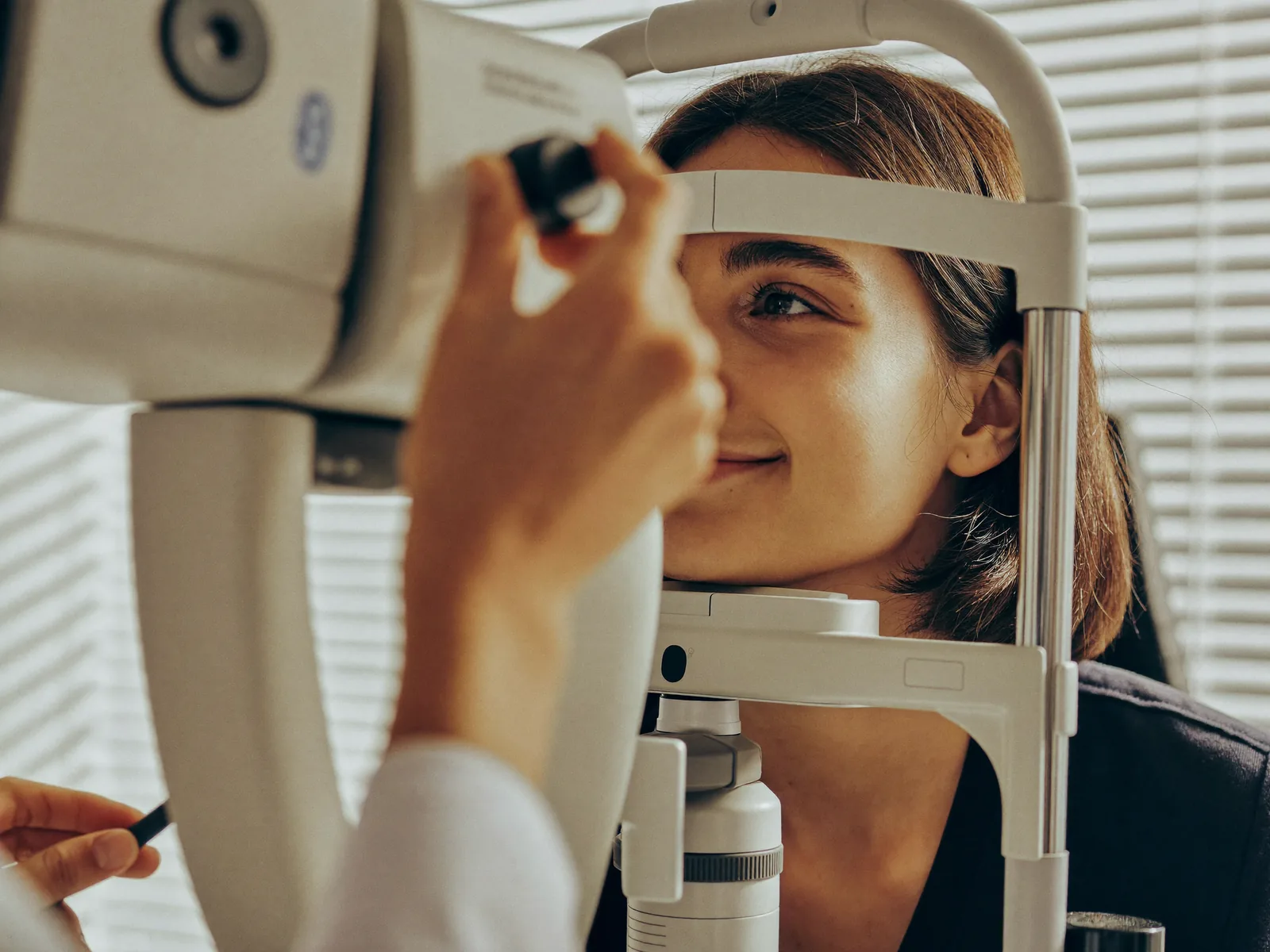


Related Pages
What Is a Full Eye Examination, and What Does It Check?
What Is a Routine Eye Examination, and How Often Should You Have One?
How Is a Glasses Prescription Measured? The Refraction Test
Why Does Blurry Vision Happen?
Laser Eye Surgery
Cataract Symptoms, Surgery and Intraocular Lens (IOL) Options
Retinal Diseases and Treatment Options
Medical Aesthetics Treatments
Contact and Appointments
Would you like to contact the doctor with a Eye Exams assessment tailored to you?
There are two ways. If you would like the doctor to know your situation before replying, complete the Eye Exams Roadmap first; if your question is short, write directly. Both reach the same team.
Your age and a short sentence are enough; no medical terms needed. What you write is only sent when you send it; this page stores no information.