
Retinal Diseases and Treatment Options
Diabetic retinopathy and AMD need regular follow-up, while a retinal tear needs urgent assessment. Compare injection, laser and surgery. Book a consultation in Istanbul.


The retina senses light and sends visual signals to the brain; common retinal diseases include diabetic retinopathy, macular degeneration, and retinal tear or detachment. The first two are chronic and need regular monitoring, while a tear or detachment needs urgent assessment. A retina examination without delay is recommended even for mild symptoms.
Written by Op. Dr. Anıl Kaya
What Is the Retina and Macula, and How Do They Work?
The retina is a thin layer of light-sensing tissue at the back of the eye, made up of photoreceptor cells — rods and cones. Signals from these cells travel to the brain along the optic nerve, forming an image. At its centre sits the macula, responsible for sharp central vision; reading, recognising faces and driving all depend on this small but critical area.
Retinal disease can affect any layer of this tissue, and symptoms depend on where the damage occurs: central (macular) damage causes blurred or distorted vision; peripheral damage narrows or darkens the visual field; and damage at the vitreous-retina interface often brings on floaters and flashes of light. Op. Dr. Anıl Kaya guides patients in Istanbul through the assessment of retinal disease; diabetic retinopathy, macular degeneration (age-related macular degeneration, or AMD) and retinal tear/detachment are seen most often.
These three follow different courses: the first two are usually slow, chronic conditions needing regular monitoring, while a retinal tear or detachment can worsen within hours to days and needs urgent assessment. Early diagnosis is decisive in limiting vision loss in most retinal disease, so a retina examination without delay is recommended even for mild symptoms. Telling these conditions apart requires a detailed retina examination assessing the retinal tissue, blood vessels and macula.
Regions of the Retina and Where Damage Occurs
Which Retinal Symptoms Mean You Should See a Doctor?
How urgently you should see a doctor about a retinal symptom largely depends on whether it appears suddenly or develops slowly. A sudden increase in floaters, flashes of light, or a curtain or shadow spreading in from the edge of your vision can warn of a retinal tear or detachment; because this can worsen within hours to days, seeing an ophthalmologist without delay is recommended.
Other symptoms — straight lines looking bent or wavy, parts of words disappearing while reading, or blurred central vision — point more towards macular degeneration and are usually noticed over weeks to months, but still warrant an examination without delay once noticed. People with diabetes need regular retina examinations even without symptoms, since diabetic retinopathy can progress silently in its early stages.
In short, sudden, fast-worsening symptoms need urgent assessment, while slower, chronic symptoms still deserve a planned examination rather than being ignored. Floaters deserve particular attention; when this symptom is harmless and when it can signal an emergency is covered separately below.
What Symptoms Occur in Retinal Diseases?
Retinal disease often progresses quietly and may go unnoticed at first, especially when only one eye is affected and the other compensates. Floaters together with flashes of light are among the symptoms patients mention most often; because this can be an early sign of a retinal tear, it needs careful assessment.
Blurred or wavy vision, straight lines that look bent, parts of words disappearing while reading, faded colours, and a dark patch or curtain in the visual field can also point to a retinal problem. Marked difficulty seeing at night, or slow adjustment from bright light to darkness, can occur too.
Whether a symptom appears suddenly or develops slowly over weeks or months is an important clue to its cause: a sudden increase in floaters and flashes needs urgent assessment, while gradually worsening blurriness more often points to a chronic process. This can vary, so a retina examination without delay is recommended whenever any of these symptoms occur — which eye, how long, and how it has changed are all useful clues at examination.

More About Eye Floaters
Floaters are small spots, threads or shadows drifting across the field of vision, usually caused by age-related changes in the eye's gel-like vitreous — most often harmless. A sudden onset of rapidly increasing floaters, or floaters with flashes of light, can signal an emergency such as a retinal tear. Whether it is a harmless vitreous change or a sign of retinal disease can only be worked out with a detailed retina examination. The page below covers its causes and when it needs urgent assessment.
What Are the Risk Factors for Retinal Disease?
Risk factors for retinal disease differ by condition. In diabetic retinopathy, the main factor is long-standing, poorly controlled blood sugar — the longer someone has had diabetes, the higher the risk — with high blood pressure, high cholesterol, pregnancy and smoking adding further risk.
In macular degeneration, age is the most important factor; genetic predisposition, smoking, high blood pressure, obesity and long-term sun exposure can also play a part, and a family history raises the risk further.
For retinal tear and detachment, the most common cause is age-related vitreous separation; high myopia, eye trauma, previous eye surgery (cataract surgery, for example) and a family history of detachment all raise the risk.
Together, these show that people over 40, those with diabetes, those with a family history of retinal disease, and those with high myopia need closer monitoring. Knowing these factors helps decide who should be followed, and how often, more than it helps prevent the disease.
What Is Diabetic Retinopathy, in Brief?
Diabetic retinopathy is an eye condition caused by damage to the retinal blood vessels from prolonged high blood sugar, and a common cause of vision loss in people with diabetes. It usually affects both eyes and may cause no noticeable symptom at first, which is why everyone with diabetes needs regular retina examinations, even without symptoms.
Over years, high blood sugar weakens the retinal vessel walls and causes leakage; in more advanced stages, vessels can become blocked or fragile new vessels can form. Treatment may not be needed early on, when monitoring and blood sugar control take priority; but as fluid build-up or abnormal vessels develop, an intravitreal injection — and sometimes laser or surgery — becomes necessary.
Untreated cases carry a risk of permanent vision loss, rising the longer diabetes has lasted and the poorer the control. Keeping blood sugar under control helps slow the condition's progression. Macular degeneration and retinal tear/detachment, which cause similar symptoms such as floaters, are other retinal diseases worth considering. Diagnosis is made through a detailed retina examination after pupil dilation, with OCT imaging used when needed.
What Are the Treatment Options for Retinal Disease?
Treatment for retinal disease is shaped by the underlying cause and stage. Diabetic retinopathy and macular degeneration are often treated with an intravitreal injection: after topical anaesthesia, a fine needle delivers a medication called anti-VEGF into the eye to control abnormal blood vessel growth and fluid build-up. Treatment is usually not a one-off — typically a series of injections a few weeks apart, followed by less frequent maintenance injections.
For a retinal tear or detachment, options include laser photocoagulation or surgery (vitrectomy or scleral buckling). Laser photocoagulation seals the area around a tear to stop fluid passing beneath the retina and can be done as an outpatient procedure; once detachment has developed, surgery becomes necessary. In some cases, pneumatic retinopexy — a gas injection that helps the retina settle back into place — may be chosen instead.
Which method is used is decided case by case from examination and imaging findings, including OCT, alongside the eye's overall structure and any other conditions. If there is an active eye infection, for example, an injection is withheld until it is controlled, and alternative approaches are considered.
Which Retinal Treatment Option Suits Whom?
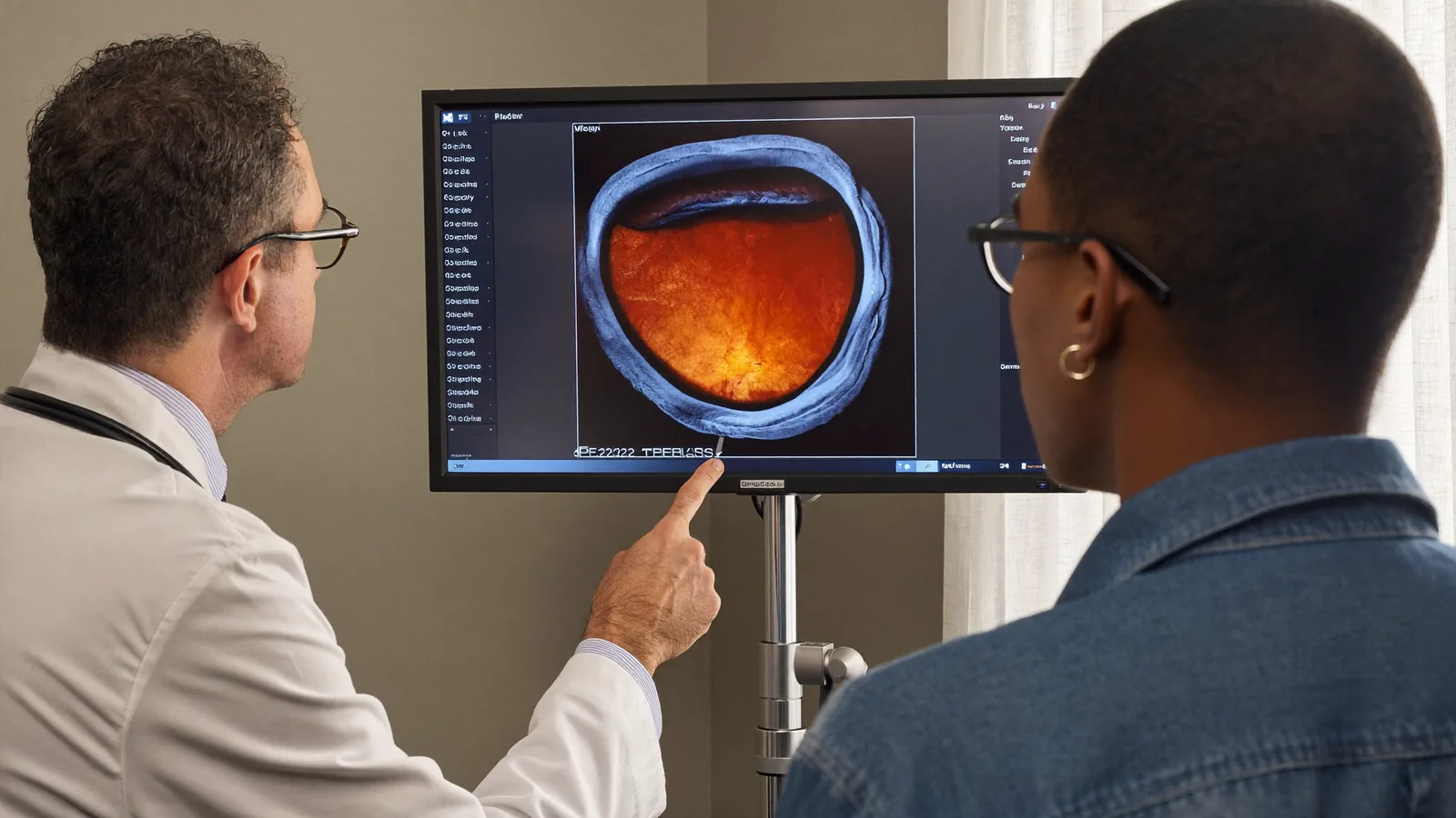
The right treatment depends on the disease's type and stage, and the eye's overall condition. In early-stage diabetic retinopathy, close monitoring alone may be enough; more advanced cases, or macular degeneration, may need an intravitreal injection. Once fluid build-up or abnormal blood vessel growth develops, treatment becomes necessary — left untreated, advanced cases carry a rising risk of permanent vision loss.
For a retinal tear, laser photocoagulation as a limited intervention may be enough; once detachment has developed, surgery can become unavoidable. Tears caught early and treated with laser can usually prevent detachment, but once it occurs, vitrectomy or scleral buckling come into play.
In some advanced or differently structured cases, an injection alone may not be enough and is considered alongside laser or surgery. This is always an individual decision made after a detailed retina examination, shaped by the eye's anatomy as much as the disease stage. The flow below summarises which option applies.
Which Retinal Treatment Suits Whom?
How Do Intravitreal Injections, Laser and Surgery Compare in Retinal Treatment?
Intravitreal injections and laser/surgical treatment serve different purposes across retinal diseases, and the right choice depends on the condition's type and stage. An injection aims to reduce abnormal blood vessel growth and fluid leakage in both diabetic retinopathy and macular degeneration; it is usually a series of injections at set intervals rather than a one-off, so repeated sessions are often needed.
Laser and surgical treatment, by contrast, come into play mainly for a retinal tear or detachment, and in advanced diabetic retinopathy to limit abnormal vessels or seal a tear; it is generally not used in macular degeneration. The choice depends on which group the condition falls into, decided individually rather than by a general rule. With very extensive damage or long-untreated detachment, the visual outcome can remain limited, and expectations need to stay realistic. The table below summarises when each is chosen.
| Intravitreal Injection | Laser / Surgery | |
|---|---|---|
| Used in diabetic retinopathy | ✓ | ✓ |
| Used in macular degeneration | ✓ | — |
| Used in retinal tear/detachment | — | ✓ |
| May require repeated sessions | ✓ | — |
How Is the Assessment Carried Out at a Retina Examination?
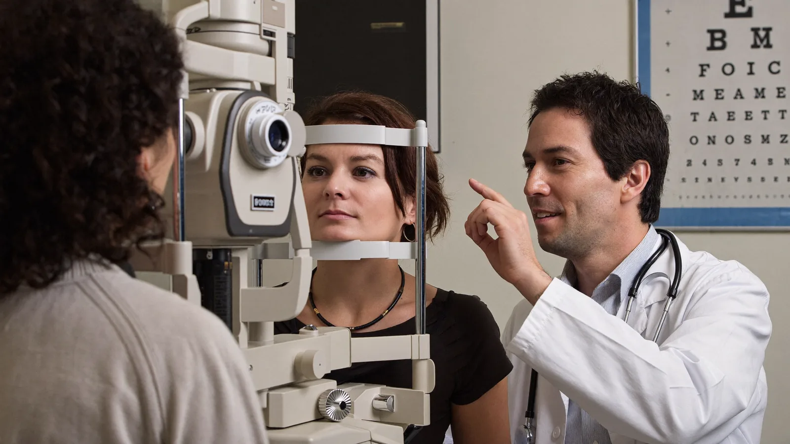
Diagnosis and treatment planning for retinal disease begins with a detailed retina examination. Drops dilate the pupil first, taking around 20–30 minutes to take effect; the doctor then uses a special lens and light source to view the retina, optic nerve and blood vessels directly.
When needed, OCT (optical coherence tomography) takes a quick, comfortable cross-sectional image of the retinal layers, requiring only that the patient rest their chin on a device. OCT reveals subtle thickening, thinning or fluid build-up, helping confirm the diagnosis and monitor treatment response. The retinal tissue, blood vessels, optic nerve head and macula are each assessed separately; bleeding, fluid, a tear, thinning or abnormal vessels can all be picked up.
Because of the dilating drops, temporary blurred near vision and light sensitivity can last a few hours, so it is best not to drive to the appointment. The diagram below summarises the main steps.
How Does a Retina Examination Proceed?
More About the Retina Examination
A retina examination, also known as a fundus examination, looks in detail at the retinal tissue, blood vessels, optic nerve head and macula. People with diabetes, those over 40, those with a family history of retinal disease, and those with high myopia should have it at regular intervals. The result can show a healthy retina, or reveal bleeding, fluid build-up or vascular changes. The page below covers who needs it, how it is done, and what the results mean.
How Does the Intravitreal Injection Process Work in Retinal Treatment?
Before an intravitreal injection, a detailed retina examination and OCT imaging clarify the type and severity of the condition. The eye is numbed with drops and the area disinfected; a fine needle then delivers the medication into the vitreous cavity through the white of the eye (the sclera). The procedure takes only a few minutes and is not usually painful, though mild pressure can occur.
Redness, stinging or temporary blurred vision can last a few days; the eye should not be rubbed, and prescribed drops should be used. Most patients return to daily life the same day or the next, though the treated eye should be protected from dirt and water for a few days, with heavy exertion and swimming postponed on the doctor's advice.
Risks include a temporary rise in eye pressure, infection (endophthalmitis), bleeding and, rarely, retinal detachment; these risks are generally low, but report any unusual symptom quickly. Sudden pain, a marked rise in light sensitivity, or sudden vision loss should prompt urgent contact with the doctor.

More About Intravitreal Injection in Retinal Treatment
Intravitreal injection is one of the most common treatments for diabetic retinopathy and macular degeneration. Carried out under local anaesthesia as an outpatient procedure, it is usually completed within a few minutes. Response varies from patient to patient; the aim is to preserve existing vision and slow progression. The page below covers who it suits, how it is done, aftercare, and its risks.
How Does the Recovery Timeline Progress After Retinal Treatment?
Recovery after retinal treatment varies considerably by method. After an intravitreal injection, most patients return to daily life the same day or the next; redness or mild discomfort can last a few days, during which the treated eye should be protected from dirt and water. The timeline below summarises this general course; the days given are approximate.
Recovery after laser treatment is also generally quick, with a return to normal activities within a few days. After surgery (vitrectomy or scleral buckling), the process can take longer — healing over weeks, sometimes with a face-down position, an eye shield, and avoiding heavy activity. Vision also changes at a different pace: an injection's effect is usually assessed within a few weeks, while how much vision returns after surgery depends on how long the detachment lasted and whether the macula was affected. The exact course becomes clear at follow-up appointments.
Recovery Timeline After Intravitreal Injection
What Should Be Done in an Emergency With Retinal Tear or Detachment?
The most typical symptoms of a retinal tear or detachment are sudden floaters, flashes of light, and a curtain or shadow spreading in from the edge of the visual field. These usually appear suddenly and can worsen over time; detachment is time-sensitive, and early intervention directly affects the chances of preserving vision.
A retinal tear or detachment can occur at any age, though risk is higher with high myopia, eye trauma, or older age. Anyone noticing this should see an ophthalmologist without delay. A limited tear found at examination can be sealed with laser photocoagulation to prevent detachment; once it has occurred, vitrectomy, scleral buckling or pneumatic retinopexy is used, chosen by size, location and duration.
Detachment usually starts at a point in the peripheral retina and, untreated, spreads towards the macula; intervention before the macula is affected offers a better chance of preserving vision.
Emergency Pathway for Retinal Tear and Detachment
More About Retinal Tear and Detachment
A retinal tear is a small tear in the retinal tissue; detachment is when fluid seeping through it lifts the retina from its normal position. This needs urgent assessment and, untreated, can cause permanent vision loss. Tears caught early can often be controlled with a limited laser treatment, while surgery becomes necessary once detachment has developed. The page below covers the symptoms, causes and emergency steps.
Does Macular Degeneration Get Better, or Does It Cause Blindness?
Macular degeneration (age-related macular degeneration, or AMD) is the gradual deterioration of the macula, responsible for sharp vision; it occurs most often after 50. It has two forms — dry and wet: dry usually progresses slowly, while wet involves abnormal blood vessels and fluid leakage, causing vision loss that becomes noticeable over weeks to months.
Whether it leads to complete vision loss depends on type and stage: central vision is usually affected, peripheral vision mostly preserved, and total loss rare. Even with treatment, fully regaining lost vision is not possible for every patient; the aim is to preserve remaining vision and slow progression.
Genetic predisposition is a known risk factor, and family history raises the risk, though age and smoking also play a part. In the dry form, regular monitoring and lifestyle measures such as stopping smoking and eating well come to the fore; these are assessed individually at examination. Diagnosis is made through a retina examination and OCT imaging, which reveals thickness changes and fluid build-up in the macula and helps distinguish dry from wet.
What Are the Risk Groups and Check-Up Frequency for Retinal Disease?
Certain groups need closer monitoring for retinal disease: people over 40, those with diabetes, those with a family history of retinal disease, and those with high myopia. A retina examination is recommended as soon as diabetes is diagnosed, even without symptoms, since diabetic retinopathy can progress silently.
Outside these groups, an annual retina examination is often enough; those with a risk factor such as diabetes may need it more often, at intervals the doctor sets. It is advisable not to drive to the appointment, since the drops can blur vision and increase light sensitivity for a few hours. Because vitreous separation can occur earlier and more often with high myopia, this group also needs close monitoring for tear and detachment.
A family history of retinal detachment, or previous eye surgery such as cataract surgery, also raises risk. Regular fundus check-ups are valuable for early diagnosis even without symptoms; the examination generally causes no discomfort and, with pupil dilation, takes around 30–45 minutes.
Frequently Asked Questions About Retinal Disease
Below are questions our patients often ask about retinal disease and its treatment, with brief answers; the information here is general, and detailed, personal guidance is given at examination.
Can retinal disease get better on its own?
This depends on the type and stage of the condition. Harmless, vitreous-related floaters can become less noticeable over time, but conditions such as diabetic retinopathy, macular degeneration or a retinal tear do not resolve on their own and need monitoring or treatment; the exact picture only becomes clear with a retina examination.
Can retinal disease cause blindness (permanent vision loss)?
Some retinal diseases left untreated, particularly advanced retinal detachment or wet macular degeneration, can cause serious vision loss. Early diagnosis and regular monitoring reduce this risk considerably; the course for any individual can only be assessed through examination.
Is macular degeneration genetic?
Genetic predisposition is one of the known risk factors for macular degeneration; people with a family history of the condition may be at higher risk. However, other factors such as age and smoking also play a part — it is not a genetic condition on its own.
How quickly does retinal disease progress?
The course varies by condition. Diabetic retinopathy and the dry form of macular degeneration usually progress slowly over years, wet macular degeneration can worsen over weeks to months, and a retinal tear or detachment can worsen within hours to days. Examination is important for telling these apart.
Are vitamin supplements helpful in macular degeneration?
In some cases, specific vitamin and mineral supplements recommended by the doctor can support slowing the rate of progression. However, this is a decision that needs to be assessed for the individual patient and the stage of the condition; rather than taking vitamins on your own initiative, it is advisable to follow the doctor's recommendation after examination.
Is surgery always necessary for retinal disease?
No. In many retinal diseases, non-surgical methods such as an intravitreal injection or laser photocoagulation can be enough. Surgical treatment usually comes up in more advanced situations, such as retinal detachment; which method is appropriate is decided from the examination findings.
How long does a retina examination take, is it painful, and which age group is at higher risk?
A retina examination generally causes no discomfort and, including pupil dilation with drops, can take around 30–45 minutes in total. People over 40, those with diabetes, and those with high myopia are the risk groups who need closer monitoring for retinal disease.
What are the most common retinal diseases?
The most commonly seen retinal diseases include diabetic retinopathy, macular degeneration (age-related macular degeneration) and retinal tear/detachment; each requires a different set of symptoms to watch for and a different treatment approach.
What are the symptoms of retinal disease?
Retinal disease can show itself through symptoms such as blurred vision, a dark patch or distortion in the visual field, sudden flashes of light, or an increase in floaters. An examination without delay is recommended whenever any of these symptoms is noticed.
How are diabetes-related retinal problems treated?
Treatment for diabetes-related retinal problems (diabetic retinopathy) depends on the stage of the condition; approaches range from an intravitreal injection to laser and, when necessary, surgical options.
What should you pay attention to in order to protect your retinal health?
Regular fundus examinations, keeping chronic conditions such as diabetes and high blood pressure under control, and staying away from smoking all play an important part in protecting retinal health.
References
- Diabetic Eye Disease — American Academy of Ophthalmology (AAO), 2024
- Detached Retina — American Academy of Ophthalmology (AAO), 2025
Book an Appointment for Your Retinal Health
If you have noticed a sudden change in your vision, floaters or flashes of light, you can book an appointment to have your retina assessed. If you have diabetes or fall into a risk group, regular retina check-ups matter even without symptoms. At your appointment your eyes are assessed, options reviewed, and questions answered. Early assessment affects treatment success in most retinal disease.
Find the Right Path for You
There are many different conditions and options in this area — browse by topic below, or answer a few questions to see the step closest to your situation.
Explore by Topic
The Basics
4 pagesDiagnosis & Methods
1 pageFind What Suits You
The tool will load shortly — or you can contact us directly.



Related Pages
How Is a Retina Exam (Dilated Eye Examination) Performed?
What Are Retinal Tear and Retinal Detachment?
What Is Intravitreal Injection (Anti-VEGF Injection) Treatment?
What Is Retinal Laser Treatment and Retinal Surgery?
What Do Eye Floaters Mean?
What Is a Full Eye Examination, and What Does It Check?
Contact and Appointments