
How Is a Retina Exam (Dilated Eye Examination) Performed?
Drops widen the pupil so the retina, optic nerve and vessels are examined, with an OCT scan if needed; vision may blur for a few hours. Book an eye examination in Istanbul.
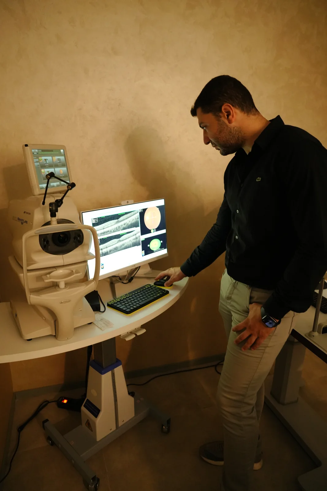

A retina exam is a close look at the retina, optic nerve and blood vessels once the pupil is dilated with drops — also called a dilated eye examination. OCT can add a cross-sectional retina image if needed. It doesn't hurt, though the drops cause blurred vision and light sensitivity for hours afterwards.
Written by Op. Dr. Anıl Kaya
What Is a Retina Exam?
A retina exam is a detailed look at the retina — the light-sensitive layer at the back of the eye where vision forms. It's sometimes called a dilated eye examination, since drops are used first to widen the pupil so the retina, optic nerve and blood vessels can be viewed directly. Many retinal diseases — including diabetic retinopathy, age-related macular degeneration (AMD), and retinal tear or detachment — are often first picked up this way. Unlike a routine eye examination, which centres on your glasses prescription and the eye's surface, a retina exam focuses specifically on the back of the eye: the retina and the optic nerve.
Quick Facts
Including the time needed for the dilating drops to work, a retina exam usually takes 30-45 minutes in total, though the examination itself takes only a few minutes. There's no cutting or injection involved, so it isn't painful — just a slight pressure when the lens comes close to your eye. People with no symptoms are usually advised to have one once a year; with a risk factor such as diabetes or high myopia, your doctor may suggest checking more often. As the drops keep working for a few hours afterwards, avoid driving yourself home that day.
What Does a Retina Exam Assess?
A retina exam checks the retinal tissue, blood vessels, optic nerve head and macula. Bleeding, fluid build-up, tears, thinning or abnormal vessels can all be picked up, and OCT adds a cross-sectional image of the retinal layers for finer detail.
The exam isn't limited to the centre of the retina — its peripheral areas are checked too, since a retinal tear often starts there. Vessel calibre, colour and course are also assessed; narrowing, blockages or leakage can be an early sign of diabetic retinopathy. The optic nerve head's colour and edge sharpness are noted too, as changes there can point to other conditions.
Some of this is already covered in a routine eye check; a suspected retinal disease calls for a more focused retina exam.

Who Should Have a Retina Exam?
Diabetic patients, people over 40, those with a family history of retinal disease, and people with high myopia are considered higher-risk and worth watching more closely. Anyone noticing eye floaters, sudden vision loss, flashes of light, or a dark patch in their field of vision should also have a retina exam without delay.
Even with no symptoms, regular retina checks are a valuable habit for early detection. How often varies from person to person; the chart below gives an approximate guide by risk group — your doctor confirms the exact frequency at your exam.
How often is a retina exam recommended by risk group?
What Symptoms Call for a Retina Exam?
Eye floaters and flashes of light are among the most common reasons for a retina exam; in some patients, they warn of a retinal tear. Blurred or wavy vision, bent-looking straight lines, and a marked drop in night vision are further reasons to get checked.
A curtain or shadow over part of your vision needs urgent assessment too, as it can point to a retinal tear or detachment. Sudden vision loss with no pain, a marked rise in flashes, or a sudden increase in floaters all call for a retina exam without delay.
These symptoms can ease off on their own at times, but that doesn't mean the issue has gone — an assessment should still be carried out.
How Is a Retina Exam Performed?
Drops are applied first to dilate the pupil, taking around 20-30 minutes to work; you may notice your near vision blurring while you wait, which is expected. Because of the drops, near vision stays blurred and light-sensitive for a few hours afterwards, so it helps to bring sunglasses. The steps below outline the general flow of a retina exam.
- 1Pupil Dilation. Drops are applied to dilate the pupil; they take around 20-30 minutes to start working.
- 2Waiting for the Drops to Work. You wait in the reception area for a short while until the drops take effect.
- 3Examination with a Biomicroscope or Ophthalmoscope. Using a special lens and light source, the doctor examines the retina, optic nerve and blood vessels inside the eye in detail.
- 4OCT Imaging, If Needed. If a finding needs a closer look, an OCT scan captures a cross-sectional image of the retinal layers; the scan is quick and doesn't hurt — you simply rest your chin on the device.
- 5Reviewing the Findings. The examination findings, along with any OCT results, are reviewed together and discussed with you.
What Do Retina Exam Results Mean?
The exam may find your retina healthy, or pick up bleeding, fluid build-up, a tear, or vascular changes. On OCT, retinal-layer thickening or thinning shows a condition's stage and its response to treatment.
Results are read alongside your general health and symptoms; one measurement isn't always enough, and follow-up exams may be needed. A healthy result means regular checks continue at your recommended interval.
A finding alone isn't a diagnosis — its type, location and size together show what it points to and what follow-up or treatment it needs. Some findings are simply monitored; others may lead to an intravitreal injection or retinal laser treatment and surgery, depending on your clinical picture.
What Conditions Can a Retina Exam Detect?
The conditions most often picked up on a retina exam are diabetic retinopathy, age-related macular degeneration (AMD), and retinal tear or detachment — each with different findings and its own follow-up or treatment approach. Less commonly, the exam can also detect retinal vein occlusion. The order below reflects relative frequency in clinical practice, not a percentage or rate.
Which findings does a retina exam pick up more often, and which are rarer?
What Are the Treatment Options If a Condition Is Found?
When a condition is found, treatment depends on its type. Intravitreal injection is often first choice for diabetic retinopathy and macular degeneration, while a tear or detachment may call for laser or surgery.
An intravitreal injection delivers medication that slows abnormal vessel growth or reduces fluid build-up, usually as a treatment programme at set intervals. For a mechanical problem like a tear or detachment, retinal laser treatment and surgery apply instead — laser seals around the tear to stop it progressing, while advanced cases may need surgery. The right approach depends on the finding and your overall eye health.
Frequently Asked Questions
Do I need to prepare for a retina exam?
Since the drops can leave you with blurred vision and light sensitivity for a few hours afterwards, avoid driving yourself to the appointment.
How often should a retina exam be repeated?
For people with no particular risk factors, once a year is usually enough; if you have a risk factor such as diabetes, your doctor may recommend checking more often.
Is an OCT scan painful?
No, an OCT scan is quick and doesn't hurt at all — you just need to look into the device.
From what age should I start having retina exams?
Without a clear symptom or risk factor (such as diabetes, high myopia, or a family history of retinal disease), a retina exam is usually added to routine eye check-ups around age 40; if you do have a risk factor, it's recommended without waiting for that age.
Is any injection or cutting involved in a retina exam?
No — the retina exam itself is entirely imaging-based and involves no injection, cutting or intervention of any kind. The only thing applied is a drop to dilate the pupil.
Is a retina exam the same as an eye pressure check?
No, these are two different measurements. Eye pressure (intraocular pressure) is measured with a separate device, while a retina exam is based on directly viewing the retinal tissue; a comprehensive eye examination often includes both together.
If a finding is picked up, does treatment start the same day?
Usually not; the finding is first assessed in detail, further imaging such as OCT is carried out if needed, and a suitable treatment plan is worked out together with you. Unless it's an emergency, this assessment is completed before any treatment begins.
References
- What Are Dilating Eye Drops? — American Academy of Ophthalmology (AAO), 2025
- What Is Optical Coherence Tomography? — American Academy of Ophthalmology (AAO), 2026
Book an Examination
If you think you're in a higher-risk group, or notice any of the symptoms above, you can book a retina exam to have your eye health assessed. Bringing your eye health history and any previous results to your appointment with Op. Dr. Anıl Kaya helps the assessment move faster. To book, or ask a question, reach us through the details on our contact page.
Find the Right Path for You
There are many different conditions and options in this area — browse by topic below, or answer a few questions to see the step closest to your situation.
Explore by Topic
The Basics
4 pagesDiagnosis & Methods
1 pageFind What Suits You
Retina RoadmapA few questions, the right next step for you


Related Pages
Retinal Diseases and Treatment Options
What Is Intravitreal Injection (Anti-VEGF Injection) Treatment?
What Do Eye Floaters Mean?
What Is Retinal Laser Treatment and Retinal Surgery?
What Are Retinal Tear and Retinal Detachment?
What Is a Full Eye Examination, and What Does It Check?
What Is a Fundus Examination and Why Is It Done?
Contact and Appointments
Would you like to contact the doctor with a Retina assessment tailored to you?
There are two ways. If you would like the doctor to know your situation before replying, complete the Retina Roadmap first; if your question is short, write directly. Both reach the same team.
Your age and a short sentence are enough; no medical terms needed. What you write is only sent when you send it; this page stores no information.