
How Is a Glaucoma Test Performed?
A glaucoma examination measures eye pressure with tonometry, scans the optic nerve with OCT and tests the visual field if needed. Book an eye examination in Istanbul.
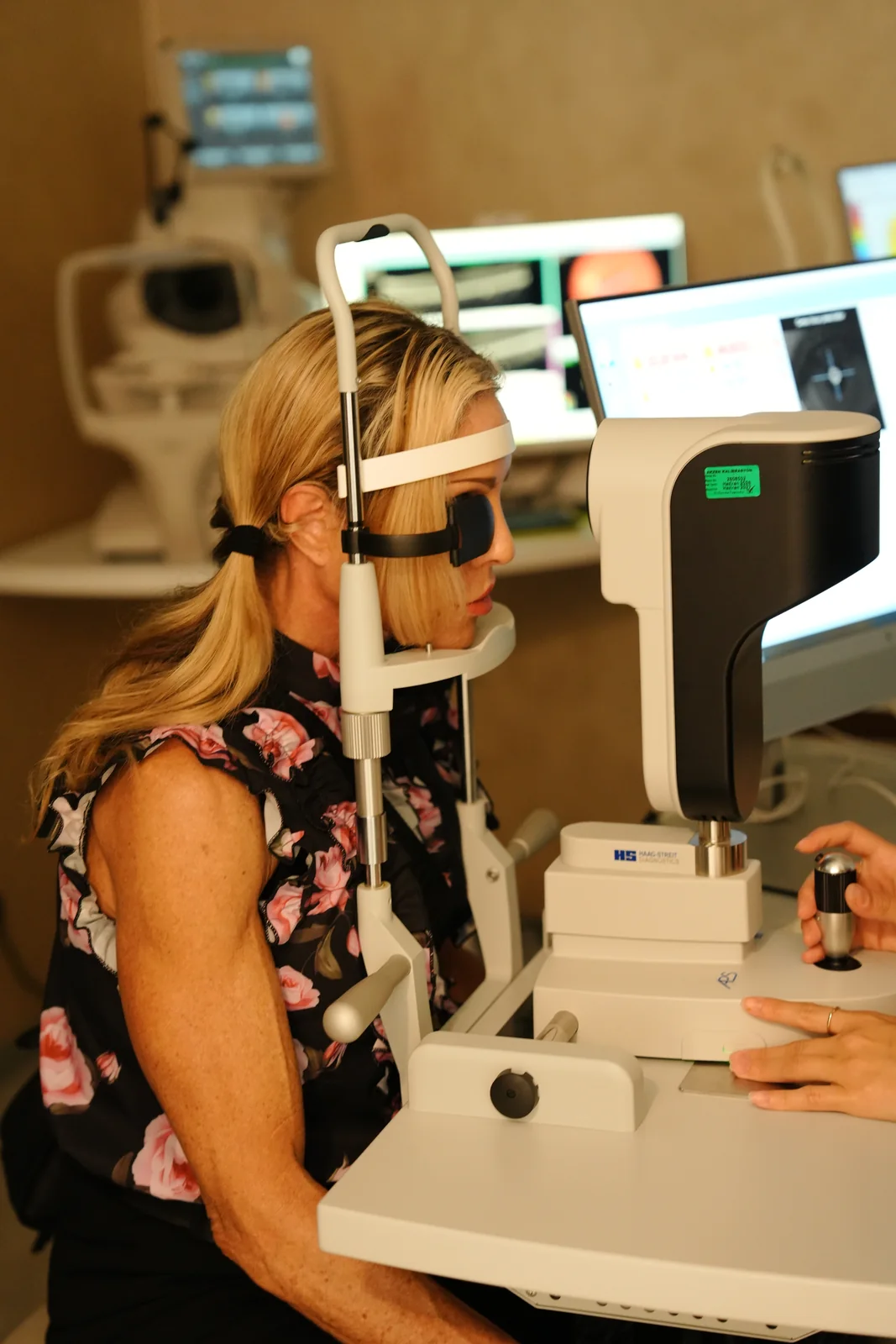

A glaucoma test is a comprehensive eye examination that assesses intraocular pressure and optic nerve health together, to diagnose glaucoma or monitor a suspected case. Tonometry measures eye pressure, OCT examines the optic disc and nerve fibre layer, and a visual field test is used when needed; corneal thickness is also recorded for accurate interpretation.
Written by Op. Dr. Anıl Kaya
What Is a Glaucoma Test?
A glaucoma test is a comprehensive eye examination that assesses intraocular pressure and the condition of the optic nerve together, either to diagnose glaucoma or to monitor a suspected case. It is not a single measurement: intraocular pressure, the structure of the optic disc, corneal thickness and, when needed, the visual field are all assessed together.
This combined approach distinguishes people with raised pressure but no optic nerve damage yet from patients with an actual diagnosis of glaucoma.
Because glaucoma usually progresses without obvious symptoms, the test aims not only to explain an existing complaint but to catch unnoticed damage at an early stage. It is therefore recommended both for people with a specific complaint and for symptom-free people at risk.
At our clinic in Istanbul, the examination is tailored by Op. Dr. Anıl Kaya to the patient's age, risk factors and any previous records.
Quick Facts
A glaucoma test is usually completed within 20 to 30 minutes; if the pupils need dilating for fundus or optic nerve imaging, it can take longer. Tonometry, a biomicroscopic optic nerve exam and, when required, OCT imaging and a visual field test are standard steps. Most tests do not typically cause discomfort; dilating drops can leave vision temporarily blurred for a few hours.
What Does a Glaucoma Test Assess?
A glaucoma test assesses three main elements: intraocular pressure, measured with tonometry; the structure of the optic disc and nerve fibre layer, examined with a biomicroscope and OCT; and, where necessary, the visual field, measured with perimetry. Corneal thickness is also recorded so the pressure reading can be interpreted correctly, since a thin or thick cornea can make the measured value differ from the true pressure.
In some patients, gonioscopy may be added to view the drainage angle directly, helping distinguish open-angle from angle-closure glaucoma. None of these measurements is sufficient alone — what matters is assessing the findings together.
Who Should Have a Glaucoma Test?
A glaucoma test is particularly recommended for people over 40, those with a family history of glaucoma, high myopia, a systemic condition such as diabetes or high blood pressure, and anyone with a previous eye injury. In these risk groups, because glaucoma can progress without symptoms, regular intraocular pressure checks matter. People with no complaints but in a risk group are also advised to have periodic eye examinations.
Recommended frequency depends on age, family history and earlier findings such as intraocular pressure; the intervals below are general guidance, and the exact frequency is set by the doctor after examination.
What Glaucoma Test Frequency Is Recommended by Risk Group?
What Symptoms Call for a Glaucoma Test?
Noticing a narrowing of your visual field, difficulty seeing objects at the edges, marked difficulty with night vision, or a tunnel-like sensation are among the symptoms that call for a glaucoma test. More on the possible causes of this complaint, and when to take it seriously, is on our peripheral vision loss page.
Sudden severe eye pain, nausea, halos around lights or sudden redness point to a different level of urgency and can be associated with angle-closure glaucoma; these should be assessed without delay.
However, most glaucoma progresses without symptoms, so regular examination is recommended for risk groups even without a complaint.
How Is a Glaucoma Test Performed?
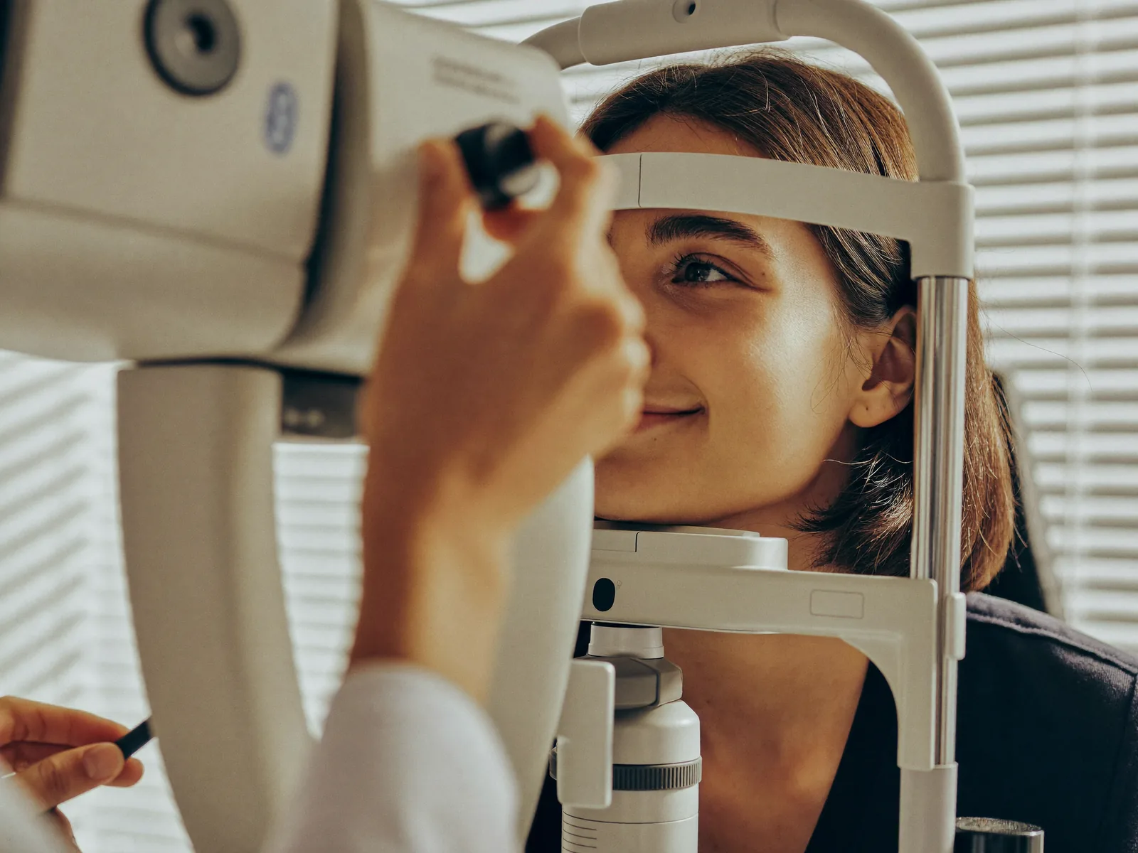
During the examination, intraocular pressure is measured first with a tonometer. The front of the eye and optic disc are then assessed with a biomicroscope; if needed, OCT measures the nerve fibre layer thickness, important for detecting early-stage damage. Corneal thickness is recorded with pachymetry.
In suspected or diagnosed cases, peripheral vision is assessed with a visual field test (perimetry). None of these tests is expected to cause pain, and most are completed in one visit; some may require dilating drops.
The steps below summarise the general flow; which are used, and in what order, can vary by patient.
- 1Intraocular Pressure Measurement. An initial assessment is made by measuring intraocular pressure with a tonometer.
- 2Anterior Segment and Optic Nerve Examination. The front of the eye and the appearance of the optic disc are examined with a biomicroscope.
- 3Corneal Thickness Measurement. Corneal thickness, measured by pachymetry, helps with the correct interpretation of the pressure reading.
- 4Optic Nerve Fibre Imaging. OCT (optical coherence tomography) examines the thickness of the nerve fibre layer at micron level.
- 5Visual Field Test If Needed. In suspected or diagnosed cases, peripheral vision is assessed with a perimetry test.
What Do Glaucoma Test Results Mean?
Intraocular pressure above normal does not, alone, confirm glaucoma; what matters is whether there is structural optic nerve damage and/or a corresponding visual field loss. If pressure is high but the nerve and visual field are normal, this is monitored as raised eye pressure or suspected glaucoma.
If optic nerve damage is found with a corresponding visual field loss, glaucoma is diagnosed and staged (early, moderate or advanced). Results are interpreted against previous records, since change over time matters more in glaucoma monitoring than a single measurement.
For patients followed as suspected glaucoma, the aim is a regular follow-up plan that catches change early, not waiting for damage to occur; a suspicious finding is a reason for monitoring, not concern.
What Conditions Can a Glaucoma Test Detect?
The most important condition a glaucoma test detects is glaucoma itself; findings help determine its stage and the appropriate treatment step. Sometimes, before a diagnosis is confirmed, only a raised intraocular pressure is found — an intermediate picture called suspected glaucoma or raised eye pressure — and the patient is monitored at set intervals. General information about glaucoma, its course and risk factors is on our glaucoma page.
Findings unrelated to glaucoma may also be noticed: early lens clouding (cataract), a retinal finding worth attention, or an uncorrected refractive error. If found, you can look into the relevant subject on our cataract, retina or laser eye surgery pages, and book a separate appointment if needed.
The grouping below reflects a general tendency, not an exact proportion; the definite diagnosis is made through the doctor's assessment.
How Common Are the Conditions Detected During a Glaucoma Test?
What Are the Treatment Options If Glaucoma Is Detected?
If glaucoma is diagnosed, or suspected glaucoma is confirmed, a treatment plan is built to bring intraocular pressure to a target level. It can start with drops and progress step by step to laser or surgery if needed. Detailed treatment information is on our glaucoma treatment page.
When drops give an insufficient response, or are unsuitable, laser options are considered; when medication and laser are not enough, or the disease has advanced, surgery is considered instead. The appropriate step depends on the pressure level, the degree of optic nerve damage and the rate of progression.
If a cosmetic eyelid finding is noticed, the patient may be referred separately for an eyelid surgery assessment, a process separate from glaucoma treatment.
Frequently Asked Questions
Is a Glaucoma Test Painful?
No, none of the tests used in a glaucoma test are expected to cause pain. Some tests may require the pupils to be dilated with drops, which can temporarily cause blurred vision or light sensitivity.
Are the Results Given on the Same Day?
In most cases, the measurement results can be assessed during the examination itself; however, the comparative interpretation of some tests, such as OCT and the visual field test, may need to be reviewed alongside previous records.
From What Age Should I Have a Glaucoma Test?
For people with no risk factors, a starting age of around 40 is generally recommended; for those with a family history of glaucoma or other risk factors, this can be brought forward. Your doctor will determine the most suitable interval for your personal situation.
How Often Should a Glaucoma Test Be Repeated?
How often it should be repeated depends on the level of risk and previous findings; patients with suspected or diagnosed glaucoma are usually followed at regular, close intervals, while less frequent checks may be enough for people at lower risk.
Do I Need to Prepare Before a Glaucoma Test?
Usually, no special preparation is required; however, if pupil dilation is planned, it is advisable not to plan on driving that day, because of the few hours of temporary blurred vision that can follow the examination.
Is a Glaucoma Test the Same as a Routine Eye Examination?
No, a routine eye examination is a broader assessment that covers visual acuity and general eye health; a glaucoma test is a more targeted examination that focuses on intraocular pressure, the optic nerve and, when needed, the visual field, aimed at monitoring suspected glaucoma or making a diagnosis.
Are My Pupils Dilated at Every Examination?
No, dilating the pupils with drops is only used when the fundus or the optic nerve needs to be examined in more detail; this decision is made by the doctor based on the findings during the examination.
What Tests Are Used in a Glaucoma Test?
Main tests used in a glaucoma test: tonometry (intraocular pressure), pachymetry (corneal thickness), a biomicroscopic optic nerve exam, OCT for nerve fibre layer (RNFL) analysis, and perimetry (visual field testing). Because OCT-based RNFL analysis can detect subtle optic nerve changes early, it is widely used in glaucoma monitoring.
In suspected cases, gonioscopy may also be used to view the drainage angle directly, helping distinguish open-angle from angle-closure glaucoma. Which test is used depends on the patient's risk level, as determined by the doctor.
References
- Get an Eye Disease Screening by Age 40 — American Academy of Ophthalmology (AAO), 2026
- Glaucoma Screening — American Academy of Ophthalmology (EyeWiki), 2026
Booking an Examination
If you think you may be at risk for glaucoma, or want a routine eye examination, you can book an appointment with Op. Dr. Anıl Kaya in Istanbul through our contact page.
Find the Right Path for You
There are many different conditions and options in this area — browse by topic below, or answer a few questions to see the step closest to your situation.
Explore by Topic
Find What Suits You
Glaucoma RoadmapA few questions, the right next step for you
Related Pages
How Is Glaucoma Treated?
What Is a Full Eye Examination, and What Does It Check?
What Is a Routine Eye Examination, and How Often Should You Have One?
Cataract Symptoms, Surgery and Intraocular Lens (IOL) Options
Retinal Diseases and Treatment Options
Eyelid Surgery and Ptosis Surgery
Contact and Appointments
Would you like to contact the doctor with a Glokom assessment tailored to you?
There are two ways. If you would like the doctor to know your situation before replying, complete the Glokom Roadmap first; if your question is short, write directly. Both reach the same team.
Your age and a short sentence are enough; no medical terms needed. What you write is only sent when you send it; this page stores no information.